When two people receive the same diagnosis of a lumbar disc herniation and one has numbness running down the leg while the other has almost no pain, the difference comes not from the name of the diagnosis but from where the nerve is compressed, how strong the inflammation is and what other problems accompany it.
A disc pressing on a nerve is seen in every patient with a lumbar disc herniation, but the factors that decide the symptoms from that point on differ from person to person. This article looks at why symptoms appear so differently and at the order in which a diagnosis is made.
Why do symptoms differ when the disc diagnosis is the same?
A disc that has been pushed out of place and presses on a nerve is common to every patient with a lumbar disc herniation, but that does not make the location and intensity of pain the same. Each nerve serves a different area, so depending on which nerve is compressed, one person has numbness on the outer thigh, another feels pulling at the back of the calf, and another has only dulled sensation on the top of the foot.
Even when the same location is compressed, the degree of pain changes again with the intensity of inflammation and with accompanying problems such as spinal stenosis or muscle tension. When inflammation is severe, even a small movement causes strong pain, but when a bulge is visible and inflammation and accompanying problems are slight, symptoms can be relatively mild, so all three have to be checked together.
- Location of nerve compression — the area of numbness and pain differs with the nerve involved, such as the outer thigh, the back of the calf or the top of the foot
- Intensity of inflammation — even at the same location, severe inflammation causes strong pain with small movements
- Accompanying problems — when spinal stenosis or muscle tension is also present, the area and pattern of pain change again
Can an MRI finding alone confirm the cause of pain?
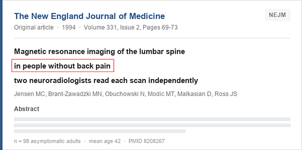
A study published in 1994 in the international journal New England Journal of Medicine is the basis for this question. When the lumbar spines of 98 adults without symptoms were scanned with MRI, disc-related abnormal findings were seen in 64 percent, and counting only discs protruding toward a nerve, such findings were still confirmed in 28 percent.
The fact that a finding is visible on an image and the fact that the finding is causing the current pain are two separate matters. In the end symptoms come before images, and without going through the process of confirming when and how the pain began, the cause of the current pain cannot be concluded from an imaging finding alone.
In what order are accurate diagnosis and treatment carried out?
When the cause differs, there is no reason for a treatment that suited someone else to suit another patient in the same way, so the candidate nerves producing the pain have to be narrowed down first. The process starts with history-taking, which asks when and how the pain began, continues with a physical examination that looks at whether the numbness changes with posture, and leads to imaging that confirms the point where the nerve is compressed.
An epidural nerve block is aimed at the point narrowed down in this way to settle the inflammation, but this procedure does not release the force pressing on the nerve itself and is a stage of dealing with the urgent problem first. Once the pain has eased, rehabilitation that rebuilds the muscles supporting the lower back has to follow so that the same spot is not compressed again.
- History — taking — confirms when and how the pain began and which posture is held for long periods during the day
- Physical examination — looks at which postures change the numbness and narrows down the location of the nerve
- Imaging — X-ray confirms the alignment of the bones, and lumbar MRI confirms the state of the disc and nerve roots