When people are diagnosed with a lumbar disc herniation, many start worrying about surgery straight away based only on what the MRI image shows.
However, what produces the pain is often a chemical inflammatory reaction around the nerve rather than how much the disc is compressed, so whether to operate is not decided from a single image.
This article looks at why MRI findings and pain do not match and why pain can return after a nerve block, and it starts with the conclusion: identifying the inflammation and the nerve location accurately, then continuing on to rehabilitation, is the key to preventing recurrence.
If an MRI shows a disc finding, is surgery always necessary?
Lumbar disc pain often comes from a chemical inflammatory reaction around the nerve rather than from physical pressure pressing on the nerve. Because irritation from inflammation, more than compression, produces the radiating pain, numbness and tingling that run down the leg, the degree of pain cannot be explained by the size of the disc bulge alone.
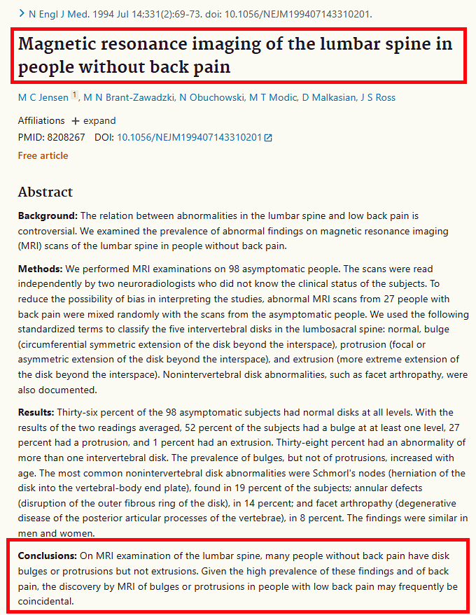
In a study that took lumbar MRI scans of 98 adults who had never had back pain, intervertebral disc abnormalities were found in 64%. Of these findings, 52% were disc bulges, 27% were disc protrusions and 1% were disc extrusions. Since such changes appeared in more than half of people without pain, it is reasonable to try treatment that controls inflammation first.
- Disc bulge 52% — the most common finding, seen in more than half of people without pain
- Disc protrusion 27% — part of the outer ring of the disc has been pushed out, and it is seen even without pain
- Disc extrusion 1% — a finding confirmed only rarely in the group without pain
Why does the pain come back soon after a nerve block?
A nerve block is not an injection given across the painful area as a whole; it is a treatment that finds the specific nerve location, among the nerve branches inside the spine, that is causing the pain now and delivers the medication there. The nerve location has to be traced back from the numb areas, the range over which the pain spreads, and which side is more uncomfortable, and if the targeted position is off, the effect drops, much like rubbing ointment on the wrist.
A nerve block only controls inflammation; it does not push a protruding disc back or make weakened muscles strong. If care stops because the pain has gone, the structure stays weak and pressure and irritation continue again in everyday movements, so manual therapy and exercise therapy need to be carried out alongside once movement becomes possible.
What should be checked when receiving treatment for a lumbar disc?
Because it is difficult to pinpoint the cause of pain from MRI findings alone, an assessment is needed that confirms the nerve location from the actual pattern of symptoms, such as the numb areas, the range over which the pain spreads and differences between the left and right sides. If a plan is made from the image alone without this assessment, the exact nerve cannot be targeted.
Even after inflammation has settled and the pain has eased, if strength and stability have not recovered, the same irritation tends to repeat and lead to recurrence. Whether rehabilitation such as manual therapy and exercise therapy is carried out alongside pain control to build strength is the standard that should be checked.
- Nerve location assessment — the process of tracing the nerve back from the numb areas, the extent of the pain and left-right differences
- Rehabilitation alongside treatment — the process of regaining weakened strength and stability through manual therapy and exercise therapy
- Checking the treatment order — confirming that inflammation control comes first and surgery is reviewed afterwards